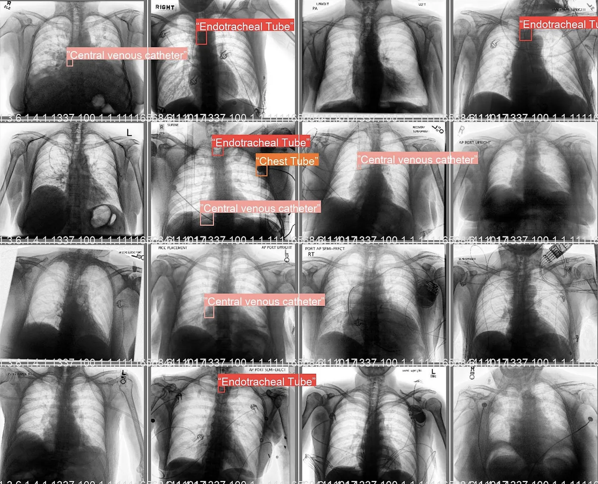
Built a two-stage chest x-ray pipeline: a multi-label classifier for device presence and a YOLOv5 detector for localization, reaching ~97% detection accuracy across heterogeneous validation sets.
Project Overview
The system preprocesses radiographs, predicts likely device classes, and then performs class-conditional bounding-box inference. Separating classification from localization reduced false positives and improved reliability under varied acquisition conditions.
Key Features
- Dual-Model Design: Multi-label screening stage + YOLOv5 localization stage.
- Detection Quality: ~97% accuracy on diverse chest x-ray validation cohorts.
- Structured Outputs: Per-class confidence scores and pixel-space bounding boxes.
- Clinical Robustness: Stable performance across different image quality and device configurations.
Technical Details
Training and evaluation were implemented in Python with dataset augmentation and threshold tuning to balance precision/recall. At inference time, the classifier gates candidate classes and the detector returns localized boxes, enabling downstream QA and faster radiology review workflows.
Internship Insights: Radiology and ResNets
During my Stanford AIMI internship, I also fine-tuned ResNet baselines for chest x-ray tasks to benchmark calibration and failure modes. Those experiments informed preprocessing and error-analysis choices for this localization pipeline.
You can read more about these insights on this Medium article.